Pharmaceutical Microbiology - Unit 4
Syllabus
Designing of aseptic area, laminar flow equipments; study of different sources of contamination in an aseptic area and methods of prevention, clean area classification. Principles and methods of different microbiological assay. Methods for standardization of antibiotics, vitamins and amino acids. Assessment of a new antibiotic.
Scroll to Download
MICROBIOLOGY UNIT-4
Designing of Aseptic Area, Laminar Flow Equipments
Study of difference of contamination in an aseptic area and methods of preventions, clean area classification.
Principles and method of different microbial Assay. Methods for standardization of antibiotics, vitamins and amino acids. Assesment of a new antibiotics
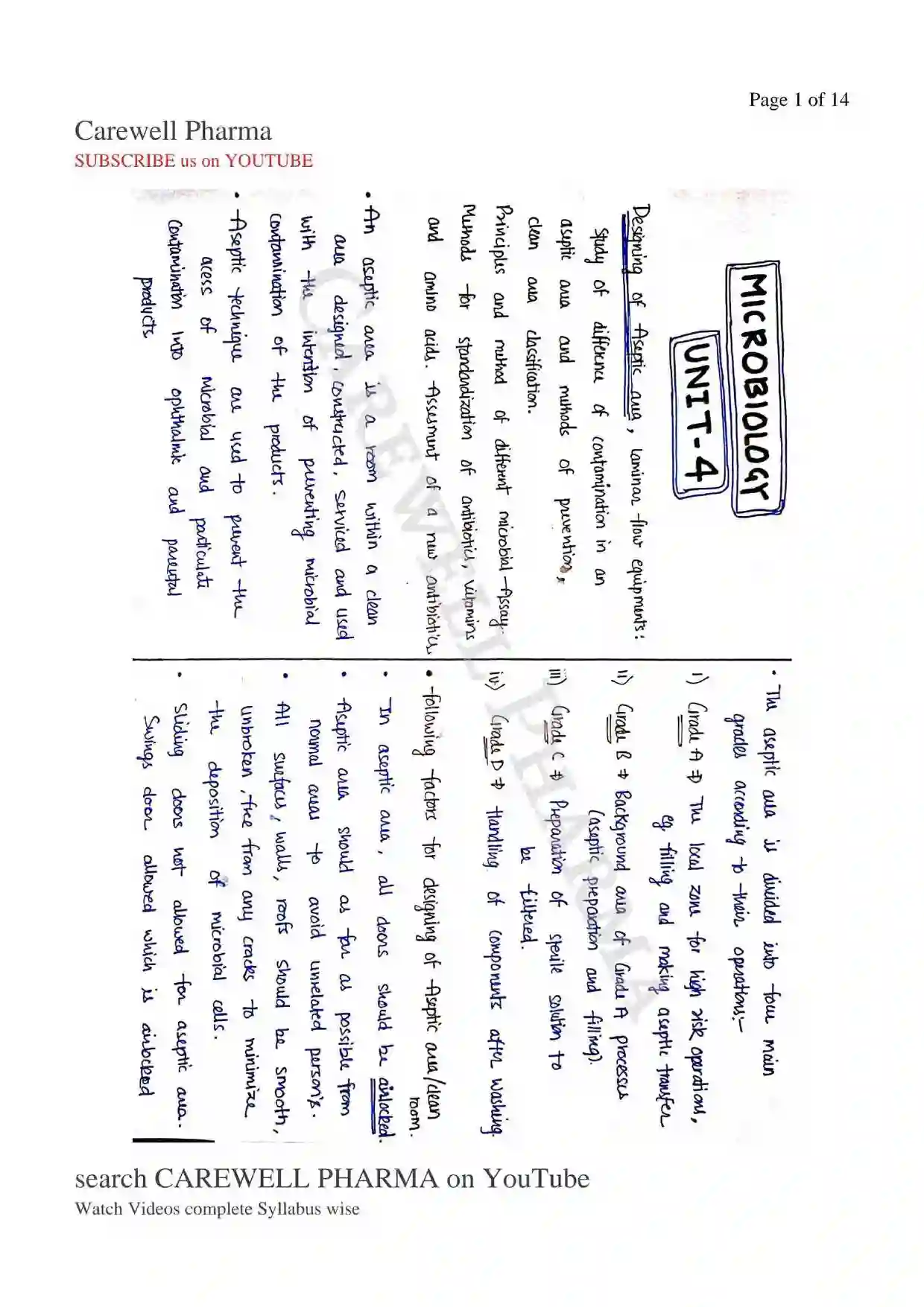
The aseptic area is divided into four main grades according to their operations:
i) Grade A The local zone for high risk operations.
eg. Filling and making, aseptic transfer
ii) Grade B Background area of Grade A processes (aseptic preparation and filling).
iii) Grade C Preparation of sterile solution to be filtered.
iv) Grade D Handling of components after washing.
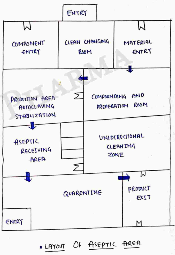
Following Factors for Designing of Aseptic Area/Clean Room.
- In aseptic area, all doors should be airlocked.
- Aseptic area should as for possible from normal areas to avoid unreleted persons.
- All surfaces, walls, roofs should be smooth, unbroken, free from any cracks to minimize the deposition of microbial cells.
- Sliding doors not allowed for aseptic area. swings door allowed which is airlocked and should be open in the flow direction of air ((+)ve air pressure to (-)ve air pressure) (+ve in room).
- Two doors can not be opened simultaneously (for prevent this warning alarm system should be there).
- All utilities (pipes and ducts etc.) should be installed in such a way that they do not create recesses, unsealed opening.
- Changing rooms should be designed as air locked, and physically separated to minimize microbial contamination.
- A filtered air supply should be used to maintain a positive air pressure relative to surroundings.
- HEPA (High efficiency particulate Air) filters should be installed to filter the air and then supplies to the aseptic rooms. which is pore size microns.
- It passes the air in aseptic area, which is free from microorganism..
Laminar Flow Equipments
Also known as Laminar air flow / Laminar flow Cabinet / Tissue Culture hood. / clean benches
Laminar flow Cabinet is an enclosed bench designed to prevent contamination of microorganism/dust at the time of Biochemical testing, performing reaction/test, also use for particles sensitive materials.
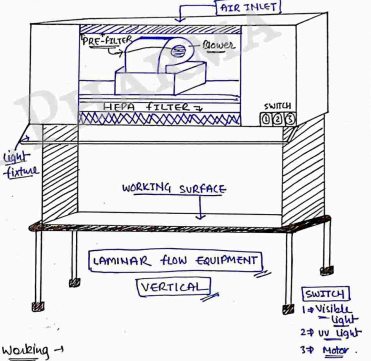
It consists of a filter pad, a fan (blower) and a HEPA filter, and one laminar air flow bench.
HEPA High Efficiency Particulate Air.
- Air is passed through a HEPA filter which remove all airborne contamination to maintain sterile Condition.
- It also consist switch for UV light, Visible light and for motor.
- It is made up of stainless steel with no gaps or joints. (for preventing bacteria).
Working
- Before starting work on laminar flow firstly on UV light for about 30 min To sterile the laminar flow.
ATTENTION
- During UV Light sterilization, stay away from Laminar flow on uv light, because it cause cancer. (maintain distance from uv light when uv is on).
- Then wiped the working surface with ethanol then use it.
- Then on the motor, the blower suck the air through the filter pad, where dust is trapped.
- After that that the prefilter air has to pass the HEPA filter where contaminating microorganism are removed.
- Sterile air flow into working area.
Types of Laminar flow Cabinet
- Horizontal Laminar air flow
- Vertical laminar air flow
Vertically Laminar Air Flow In this, air flow from top to downward (vertically). Blower (fan) is fitted upward of working area with HEPA filter.
Horizontal Laminar Air Flow
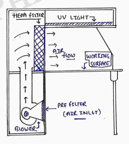
Blower is fited downward and Hepa filter on front. In this, air flow from back to front (horizontally).
Source of Contamination & Their Prevention
Proper uniform of aseptic area with fully cover body. there are various sources of contamination in aseptic area.
i) Atmosphere
ii) Operator
iii) Raw Material
iv) Equipment
i) Atmosphere → Air is the major source of contamination as it contain dust particles, microbes, spores etc.
- It may be reduced by chemical disinfection, UV Light, filtration and gaseous agent. and also by airlocked doors..
ii) Operator → The skin, hair and clothing of the operator are potent source of microbial contamination.
- Operate do any manual contamination, may be due to lack of training or focus. And operator may be skilled (trained).
iii) Raw Material → It account for a high proportion of microbial contamination into pharmaceutical products. (water is the major source).
- It may be due to improper sampling, inadequate storage condition etc
- It may be reduced by Heat treatment, filtration, and other sterilisation process in raw material. Water may be treated by UV light.
iv) Equipment → The equipments and Utensils used in processing, holding, transferring and packaging are also common source of contamination of aseptic area.
- It is due to improper design, material leads to corrosion, improper cleaning, lack of sterilization etc-
- It may be reduced that operator is in sterilized or disinfected by heat, gaseous agents or chemicals. some are also sterilized by steam under pressure (autoclave).
CLEAN AREA CLASSIFICATION
Clean room
- A cleanroom is defined as a room in which the concentration of airborne particles is controlled. (It may be controlled by HEPA filter and airlocked entry system).
- It established by measurement of the number of particles (airborne) with size or greater, that are contained in (1 cubic foot) of Sampled area.
- According to European Community guidelines it classified into Grade A, B, C and D.
- According to US Federal Standard Association classification of clean room
- Class 100 (standard applicable to aseptic areas).
- Class 1000
- Class 10000
- Class 1,00,000
- This classification is used in pharmaceutical industry.
- Class 100 means that not more than 100 particles per cubic foot of size or greater, shall be found in measured area.
- Same other classes measured according to point 2.
MICROBIOLOGICAL ASSAY
Microbiological Assay
- Also called as microbial assay.
- A microbiological assay defined as quantitative or qualitative determination of chemical compounds (antibiotics, vitamins and amino acids) with the use of microorganism.
- It is necessary to assay antimicrobial agent, for determination of potency of a drug in animals or man and monitoring or controlling antimicrobial chemotherapy.
- Many therapeutic agents, which either inhibit the growth of the microorganism (antibiotics) or are essential for their growth (vitamins & amino acids) can be standardised by microbial assay. Relative Potency used to express the biological activity of a sample preparation compared to a standard preparation.
- Microbiological assay of antibiotics (method for standardization of antibiotics)
It is based upon a comparison of the inhibition of growth of micro-organism by measured concentration of the antibiotics under examination (test antibiotics) with the known concentration of a standard known antibiotic preparation.
Two general methods are usually employed,
- Cylinder-plate or Cup-plate method
- Turbidimetric or tube assay method
Preparation for antimicrobial assay
- preparation of media & buffer solutions.
- Selection of microorganism
- preparation of test & standard solutions of antibiotics
Microorganism used in antibiotics assay:- Bacillus pumilus, Staphylococcus epidermis etc.
Method 1 (Cylinder-Plate OR Cup-Plate Method)
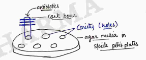
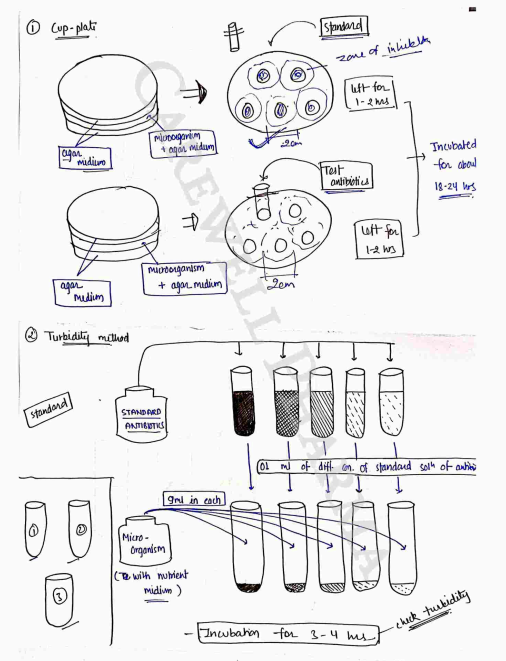
- This method depends upon the diffusion of an antibiotic from a vertical cavity or a cylinder, through a solidified agar layer in a petri-plates.
- The growth of test microorganism is inhibited entirely in a circular area or zone around the cavity/cylinder containing a solution of antibiotic.
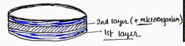
- Three layered plates are prepared for this method.
- First & third (bottom) layer is prepared of agar medium (without micro-organism).
- Second layer of agar medium (with microorganism) was prepared. (thickness - 4mm).
- Now, the holes are made in agar media by using cork borer (equal size of holes) and antibiotic solutions are filled in those holes with the help of micropipettes.
- Solution of known conc of the standard preparation and the test antibiotics are prepared in appropriate solution (acc to IP).
- These solutions are added in sterile cavities or cylinders prepared in agar medium separately (separate for test and standard solution of antibiotic).
- The plates are left standing for 1 to 2 hrs at room temp. or .
- All plates are then incubated for about 18-24 hrs at temp. (acc to IP) (acc. to bacteria, & antibiotics).
- The diameter/areas of the circular inhibition zones produced by standard and test antibiotics solution are accurately measured.
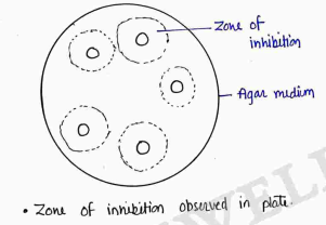
Zone of inhibition observed in plate.
- The graph which relates zone diameter to the logarithm of the concentration of antibiotics is plotted, and the unknown concentration of test antibiotics is calculated.
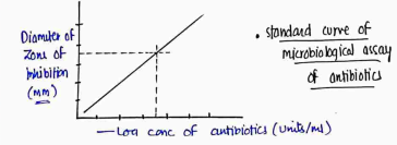
- Method 2 (Turbidimetric OR Tube assay Method)
- This method depends upon the growth of a microbial culture in a uniform solution of the antibiotics in a fluid medium that is favourable to its rapid growth in the absence of the antibiotics.
- Here, turbidity shows the microbial growth.
- It has a shorter Incubation periods for the growth of test microorganism (3 to 5 hours).
- This method is not recommended for cloudy or turbid preparations.
- Five different concentration of the standard solution are prepared by diluting the stock solution, for making the standard.
- A median concentration is selected and the test sample of the antibiotics solution is adjusted by dilution to obtain approx this concentration.
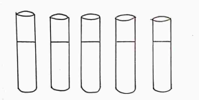
- One ml of each concentration of the standard solution and of the sample solution (test antibiotic sol^n) are placed in each of the tubes in duplicate.
- To each tube, 9 ml of nutrient medium (with test microorganism is added).
- At the same time, three control tubes, one containing the inoculated culture media, second treated with 0.5 ml of dil. formaldehyde solution (blank) and third containing un-inoculated culture media are prepared.
- All the tubes are placed in an incubator at the specific temp. (approx. $37^\circ C$) for 3 to 4 hrs.
- Now compared the turbidity of test with standard.
Microbiological assay of Vitamins
- Vitamins are important growth factor needed for growth and multiplication of microorganism, they are very sensitive to small amount of growth factors.
- It is the ability of these test microorganism to synthesize the factor being assayed that forms the basic of microbiological assay of Vitamins & Amino acids.
- Such test microorganism used for assaying the water soluble vitamins as Vitamin , Vitamin etc..
- Such test microorganism used as Lactobacillus leichmannii etc..
Assay of Cyanocobalamin (Vitamin $B_{12}$)
i) Titrimetric Method
ii) Turbidimetric Method
i) Titrimetric Method
- Take 10 clean test tube and fill 0 ml, 0.5, 1.0, 1.5, 2.0, 2.5, 3.0, 4.0, 4.5, 5.0 respectively of standard Vitamin sol^n ($0.01 - 0.04~ng/ml$). (Standard sol^n)
- Now add 5 ml Basal media stock sol^n.
- then make upto 10 ml by using water.
- Take 4 clean test tube and fill (add) 1ml, 2ml, 3ml, 4ml respectively of test solution to be assayed. (Test sol^n)
- Now add 5 ml Basal media stock sol^n to each test tube and make upto 10 ml by using water.
- Now sterilize all tubes (standard + test) in autoclave at for 5 min, then cool all test tubes upto room temp and inoculate with one drop of inoculum (test microorganism - lactobacillus...).
- Incubate the tubes for 64 to 72 hrs at temp within .
- Now, titrate the content of each test tube (standard + test sol^n). with NaOH by using 0.1% w/v bromothymol blue as an indicator or by electrometrically (converts to green color).
- Determine the average value of titration for both (standard & test).
- Now plot the graph shown below. for both.
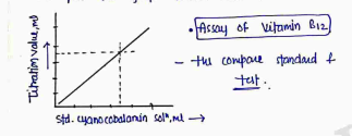
- Then compare standard & test.
ii) Turbidimetric Method
- Use device Photoelectric colorimeter.
- Apparatus, reagent and procedures are same as titrimetric method but in this includes two more test tube which is uninoculated blank tube (no standard + no test sol^n + no microorganism).
- Incubate all test tube at for 16-24 hrs.
- Now. By using 'uninoculated blank tube' adjust the transmittance at 640 nm to 100% in the photoelectric container.
- Throughly mix the contents of each tube and record the transmittance reading.
- Now plot the graph by considering transmittance value against the corresponding level of standard cyanocobalmin sol^n.
- Draw a smooth curve and calculate the concentration of the test sol^n of cyanocobalmin.
Assessment of New Antibiotic (Minimum Inhibitory Concentration) MIC
- It is the lowest concentration of antimicrobial compound found to inhibit the growth of a particular test microorganism.
- It may applied to acess new disinfectants, antiseptics, preservatives and antibiotics.
- Usually expressed in terms of or units/ml.
- MIC of different antibiotics (antimicrobial) compounds for the growth in the form of turbidity.
- May be determined by the Liquid dilution method or Solid dilution method.
1. Liquid Dilution Method (Test Tube Method)
- Take ten clean test tube and add (0.5, 1.0, 1.5, 2.0, 2.5, 3.0, 3.5, 4.0, 4.5, 5.0) ml respectively of test chemical.
- Take another two test tube, one is un-inoculated (without chemical & test microorganism) and other is control test tube used to check the suitability of the medium for growth of microorganism (without chemical).
- Now add 5 ml double-strength medium in all test tube.
- then un-inoculated tube (except un-inoculated tube).
- Now, make volume upto 10 ml by using sterile water.
- Now incubated all test tube at for 2-3 days.
- After incubation, all test tube are examined for growth in the form of turbidity.
- Now, Minimum inhibitory concentration (MIC) is calculated.
2. Solid Dilution Method
- Test chemical is first mixed with into molten agar, then poured into petri plates.
- After solidification, the inoculation inoculum is spread on the surface of agar medium.
- All plates are incubated at for 2-3 days.
- After incubation, all plates are observed and the MIC of test chemical is calculated.