Pharmaceutical Microbiology - Unit 1
Syllabus
Introduction, history of microbiology, its branches, scope and its importance. Introduction to Prokaryotes and Eukaryotes Study of ultra-structure and morphological classification of bacteria, nutritional requirements, raw materials used for culture media and physical parameters for growth, growth curve, isolation and preservation methods for pure cultures, cultivation of anaerobes, quantitative measurement of bacterial growth (total & viable count). Study of different types of phase constrast microscopy, dark field microscopy and electron microscopy.
Scroll to Download
MICROBIOLOGY UNIT-1ST
Micro + Bio + Logy $\downarrow$ Small + Living + Science.
Microbiology
Is the branch of science in which we Study about the small living organism which less than 1 (micron) which we can't see with naked eye. we use microscope to see them.
Eg : Viruses, Bacteria, fungi, Algae etc- We Study about them
History of Microbiology
- Aristotle Father of Biology - Living <......> Non-Living.
(Many living, we can't see). - Roger Becon 13th century - Disease (Anything which enter in body make sick us)
- Fracastorius 1544 - Communicable disease (those disease which spread through communication).
- Louis Pastuer Father of Microbiology - Introduced sterilization techniques. - pasteurisation of milk - 30min (milk) all bacteria kill.
- Lord Joseph Lister Father of Antiseptic Surgery - He instructed surgeons under his responsibility to wear clean gloves and wash their hand before and after operation with 5% carbolic acid soln.
- Alexender flemming First Antibiotic - Penicillin (1928) Pennicilium notatum (made from)
Branches
- Virology → Study of viruses
- Bacteriology → Study of Bacteria
- Protistology → Study of Protists
- Mycology → Study of fungi
- Parasitology → Study of parasites
- Acellular (cell not complete) eg. Viruses.
- Cellular →
- Prokaryotic eg Bacteria,
- Eukaryotic eg fungi, Algae, Protozoa
Scope & Its importance.
1) Medical → Deals with diseases of human & animals.
2) Immunology → Study of the Immune system that protects the body from pathogens.
3) Agricultural Microorganism → Impact of microorganism on agriculture.
4) Industrial Microbiology → Using microorganism to make products such as antibiotics, vaccines, steroids, alcohols etc-
5) Food & Dairy Microbiology → Use microorganism to make food such as cheeses, yogurts, pickles, beer, etc.
Classification
1) Acellular microorganism → Those in which cell does not developed properly.
Eg → Virus.

- Cellular Microorganism → Those in which cell was developed.
Prokaryotes: Those cells in which cell membrane developed, but Nucleus membrane does not developed, also some organelles not developed. $\rightarrow$ eg Bacteria, Archaeobacteria
Eukaryotes: Those cells in which, cell membrane, Nucleus, their organelles are fully developed. eg fungi, Algae, Protozoa, Nematoda.

Bacteria → It forms a large group of unicellular prokaryotic that do not contain a nucleus & other membrane bound organelles.
- Archaeobacteria → Ancient bacteria
- Eubacteria → True bacteria
Structure of Bacteria
On Ultra-microscopic view it look like as small capsule.

Parts of Bacteria
- Capsules
- Cell wall
- Cell Membrane
- Flagella
- Cytoplasm
- Nucleoids
- Inclusion body
- Mesosomes
- Ribosomes
- Pilli
1) Capsules
- Outermost thick and slippery structure and rigid and flexible.
- Composition [98% water + 2% glycoprotein].
- Basis of thickness
- Macro Capsule → > 0.2$\mu$
- Micro Capsule → < 0.2$\mu$
- It is identity of bacteria and decide its virulence (घातक).
- It gives protection to the bacteria, also give shape and size to bacteria, also prevent from phagocytosis.
- It helps in attachment and repulsion with other bacteria.
- It causes disease in human body.
2) Cell Wall → It is also thick structure, made up with peptidoglycan layers.
- We can Identify bacteria as gram (+) or gram (-) on the basis of cell wall.
- Gram (+) → 7-8 nm thickness of peptidoglycan.
- Gram (-) → 20-80 nm thickness of peptidoglycan.
- Provide protection shape and identification to the cell bacteria.
3) Cell Membrane → Thin layer in prokaryotic cells or bacteria.
- Made up with phospholipid bilayer.
- It act as semi-permeable membrane.
4) Cytoplasm → Less develop Nucleus. so it is called Nucleoid.
- Nucleoid contain, 60% DNA, 30% RNA, and 10% protein.
5) Inclusion Body → It stored food or food of bacteria is stored in the form of inclusion body.
6) Ribosomes → It helps in protein synthesis in bacteria.
- In bacteria 70s types of ribosomes present.
8) Mesosomes → It is inside in cell wall slightly. it is extra chromosomal DNA.
- Helps in cellular Respiration.
9) Flagellum → Long thick hair like structure. 15-20 nm long and 2-5 nm diameter.
- It works like as antenna, which gives signal to bacteria for food, danger, locomotion etc-
- Made up by flagellin protein.
10) Pilli → It is small thin 8-10 hair like structure.
- Helps in attachment of bacteria with other bacteria.
- Also helpful in transfer in genetic material.
Morphological classification of Bacteria
On the basic of morphological, bacteria classified into six major groups.
- Bacteria
- True Bacteria
- Spherical [Cocci/Coccus] $\rightarrow$ Monococcus $\rightarrow$ Diplococci $\rightarrow$ Tetradcocci $\rightarrow$ Streptococci $\rightarrow$ Sarcinococci $\rightarrow$ Staphylococci
- Rod (Bacillus) $\rightarrow$ Monobacillus $\rightarrow$ Diplobacillus $\rightarrow$ Tetrabacillus $\rightarrow$ Streptobacillus $\rightarrow$ Sarcinobacillus $\rightarrow$ Staphylobacillus
- Pseudo/False Bacteria
- Actinomycetes
- Spirochaetes
- Mycoplasm
- Rickettsiae
- Chlamydiae.
- True Bacteria
(1) True Bacteria → These are real bacteria and their shape is same as bacteria.
- Cocci/Coccus → These are spherical or oval shaped. these are further classified as:
- Monococcus : In which cocci is in single form.
- Diplococcus : In which cocci is in pair.
- Tetradcoccus : In which cocci is in group of four.
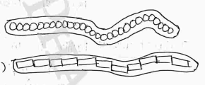
- Streptococcus : Cocci is in chain form.
- Sarcinacoccus : Cocci is in group of eight or in the form of cube. Cube form
- Staphylococcus : Cocci in grape-like clusters. Grapes like cluster.
- Bacilli / Bacillus → These are rod shaped bacteria, these are further classified as:
- Monobacillus : In which bacilli is in single form.
- Diplobacillus : In which bacilli is in pair.
- Tetrad Bacillus : In which bacilli is in group of four.
- Strepto-Bacillus : Bacillus is in chain form.
- Sarcinabacillus : Bacillus is in group of eight or in the form of cube.
- Staphylo Bacillus : Bacillus is in the form of grapes like clusters. cluster form.
(2) False/Pseudo Bacteria → These are those bacteria, which are actually a bacteria, but their shape is different from bacteria.
- Actinomycetes → (Actin-ray, Mykes - fungus)
- These are rigid organism like true bacteria.
- Resemble fungi in that they exhibit branching and tend to form filaments.
- Basically it is gram (+).
- Mostly present in soil.

Spirochaetes → These are relatively longer, slender non-branched microorganism of spiral shape.
- Basically it is gram (-).
- Length 3 to 500 (micron).

- Mycoplasm → These are in oval shaped and these are lack in rigid cell wall (cell wall lacking), and cell membrane not developed properly.
- May be coccus or bacillus, but double membrane present. [Smallest bacteria]
- Look like as virus.
- Some organelles and DNA-RNA present.
- Caused disease in Animals.

- Rickettsiae → These are look like as filaments in which no branched & no chain present.
- These are non-motile (no movement).
- Basically it is gram (-). its diameter is 0.1-0.4 (micron).

- Chlamydiae →
- It is oval-shaped.
- Peptidoglycan + protein are present in it.
- It is mostly responsible for the disease in human eye.

CULTURE MEDIA
That media (medium) in which bacteria is invite to grow. Culture media contain all essential material which is required for growth and development.
Why we need culture medium ??
- We need this for study about bacteria for any medical helps.
- Different types of bacteria need, different types of culture media for growth.
- Culture media is divide into three Category:-
Culture Media
Based on Consistency Solid $\rightarrow$ Semi-Solid $\rightarrow$ Liquid
Based on Composition Synthetic $\rightarrow$ Non-Synthetic
Based on application Basic C Medium $\rightarrow$ Enriched C Media $\rightarrow$ Selective C media $\rightarrow$ Enrichment culture media $\rightarrow$ Differential Culture media $\rightarrow$ Transport Culture media $\rightarrow$ Anaerobic culture media $\rightarrow$ Assay culture media
1) Based on Consistency
It is of these types, Solid, semi-solid and liquid. And, Agar is important for this. It is a solidifying agent.
- Eg. Nutrient Agar + Agar (1.5-2%) Solid medium
- Agar (Less than 0.5%) Semi Solid
- Agar (Absent) Liquid. eg. fluid thioglycollate broth.
- Nutrient broth containing 0.5% agar into different media.
- So, Bacteria needs depend upon the quality or quantity of material which is used to prepare culture media.
2) Based on Composition
material which is used in this is Carbon source, Nitrogen source, water, Energy source, growth factors and salts etc.
Synthetic (Highly pure) → In which we know the composition of ingredients. eg. Peptone water (1% peptone + 0.5% NaCl in water).
Non-Synthetic (Impure) → In which we don't know about the composition of ingredients. eg. Yeast extract broth etc...
3) Based on Application
In which we create culture media according to need of bacteria on bases of applications of bacteria.
i) Basic Culture Medium → It is applicable for all bacteria, because it contain all types of essential material, so in which all types of bacteria grow.
ii) Enriched Culture Media → It is for those bacteria, which need extra nutritional material (extra material blood, serum etc-) for extra growth...
iii) Selective Culture Media → These are specially designed culture medium made for special or any one bacteria.
iv) Enrichment Culture media: It is designed for some specific type bacteria and in the media, antibiotic is used for reduce unwanted bacteria so that special type of bacteria grow easily.
v) Differential Culture Media (Liquid medium) → In which metabolic dye is used to identify the different colonies by different color (dye is used for color) eg. Blood Agar.
vi) Transport Medium → It is used when culture medium is made for long time or not used frequently. And it is prevent from drying.
- It is used because, in culture media bacteria is died in 1 or 2 days, but if we need this after 15-20 day or after many days.. so we prepare this transport culture media.
vii) Anaerobic Culture Media → It is used for those bacteria, which does not required Oxygen. And in which we use Hemin or Vit. K and bacteria will grow easily.
Eg. Robertson's cooked meat medium.
viii) Assay Medium → This culture media is used for study purpose, in which we have to know about the concentration of material which is used to make culture media.
- It is right, perfect or not.
Nutritional Requirement:- FOR BACTERIA
All living beings required nutrient for their growth. So, Bacteria also need this for growth.
i) Essential Elements (Major Macro Nutrients) → These elements are important for bacteria and required in large amount.
Carbon, Hydrogen, Oxygen, Nitrogen, Phosphorus, Sulphur etc-
Hydrogen & Oxygen → These are made available from water added to culture media.
Carbon → Carbohydrates is the principal source of carbon which is degraded by bacteria - either by Oxidation or by fermentation.
Nitrogen → It is the main source of protein and nucleic acid for bacteria and its main source is ammonia.
Water → Moisture is an essential requirements for bacterial growth as 80% of the bacterial cell consists of water.
Sulphur and Phosphorus → Sulphur forms parts of structure of several coenzymes and cysteinyl and methionyl side chain of protein. Phosphorus required as component of nucleic acids, ATP, Coenzymes, NAD and flavins.
ii) Mineral Sources → These are Potassium, Calcium, magnesium, iron, copper, cobalt, manganese and zinc required in little amount for enzyme functions. and it can be provided in tap water or as contaminants of other medium ingredients.
iii) Energy Sources: Carbon dioxide and Energy is important for cellular metabolism and it can required by all bacteria and much provided by atmosphere to bacteria. source depends upon the nature of bacteria.
Aerobes → required oxygen for growth. (obligate aerobes) eg. Pseudomonas, Bacillus.
Anaerobes → which grown only in absence of oxygen. eg Clostridium, Bacteroids.
Autotrophic Bacteria → Capable to synthesized their own food.
- Photosynthetic → Use sunlight as a source of energy.
- Chemosynthetic → Sulphur as a source of energy. without photosynthetic pigments. they use chemical.
Heterotrophic Bacteria → Unable to synthesized their own metabolites.
- Photoheterotrophic → They obtain Carbon from organic compound, made by other organism.
- Chemo-heterotrophic → They take their food from another plants and animals. they obtain both energy and carbon from organic compounds.
iv) Growth Factor for Growth → Bacteria need Purine & Pyrimidine, much important for formation of DNA/RNA and also for cell division. And also required some amino acid and protein and Hormones for growth.
v) Vitamins → In many cases the growth factor are identical with vitamins of B group - thiamine, Riboflavin, Nicotinic acid, pyridoxin, folic acid and Vit B12.
Raw Material Used for Culture Media
That material, which are used in making of culture media.
I) Water
II) Peptones
III) Meat extract
IV) Agar
V) Yeast extract
I) Water
- In bacteria 70-80% of water is present and it used as a vehicle for flow of nutrients.
- So, Water is necessary for preperation of culture media. But, distilled water is prefered to use, because tap water contain calcium and magnesium, which may be react with phosphate present in peptones, and may be inhibits the growth of bacteria.
II) Peptones
- It is partially complex mixture of partially digestive protein obtain from meat, heart muscle, fibrin, soya meal etc.
- It is hygroscopic in nature (which attract water from its surroundings).
- It contain certain growth factor, imp. constituents like protease, aminoacids, magnesium, potassium which supply nitrogenous material and also act as a buffer.
III) Meat Extract
- It contain gelatin, protease, creatine, purines and growth factor etc which is essential for bacteria and it prepared from fresh lean meat by hot water extraction process.
IV) Agar
- It is important material for culture media, and it is used for preperation of solid media.
- It is long chain polysaccharide obtained from seaweed's algae.
- It act as a good solidifying agent.
- It is easily available.
- It has no nutritional value in media.
- Microorganism cannot eat and destroy it.
V) Yeast extract
- It consists of the cell contents of the yeast without the cell walls, it contain carbohydrates, amino acid, growth factor, inorganic salts. Used mainly as a source of vit. and substitute for meat extracts.
Physical Parameter for Growth
There are many physical condition, which are essential for bacterial growth or it affect the growth of bacteria.
- Temperature → It is one of the important factor that for growth.
Optimum growth temp - That temp, bacteria grow rapid and maximum.
Different bacteria grow at different temp.
- The highest temp at which microbes shows growth is known as maximum growth temp.
- The lowest temp at which microbes shows growth is called minimum growth temp.
Based on temp. they classified
I) Psychrophiles ($0^\circ C - 15^\circ C$)
II) Mesophiles ($20^\circ C - 40^\circ C$)
III) Thermophiles ($45^\circ C - 70^\circ C$) or may be more.
- pH → All bacteria grow in a certain pH range.
- Acidophiles (1 - 6.5)
- Neutrophiles (6.5 - 7.5)
- Alkalophiles (7.5 - 14)
- Gaseous Requirement → The principal gas that affects growth are and .
- Aerobic bacteria Required for growth.
- Photosynthetic and Chemosynthetic bacteria Required for growth.
- Osmotic Pressure
- Bacteria can tolerate osmotic variation due to mechanical strength of cell wall.
- They can grow in media with varying contents of salts, sugar and other solutes.
- Light
- Bacteria are Light sensitive (sensitive to UV Light, direct light (sunlight) and other radiations.
- So, Darkness is usually favorable for growth of all microbes.
Growth Curve
Bacterial Growth Curve
When a graph plot between the growth of bacteria and time phase is called growth curve.
The resulting curve has four distinct phases
I) Lag phase
II) Log phase
III) Stationary phase
IV) Decline phase (Death phase).

I) Lag Phase : In this phase, their are no growth of bacteria, because culture media is just prepared, so upto 2-3 hour their are no growth of bacteria, only nutrition medium present.
II) Log Phase : In this phase, their are maximum growth of bacteria, because after the germination of bacteria, they reproduce in fast rate and bacteria increases, and their are nutritions are more than the bacteria, so bacteria grow maximum.
- Also called as exponential phase.
III) Stationary Phase : In this phase, Growth of bacteria stop, and it may be stationary, because no. of birth of bacteria is equal to the no. of death of bacteria.
- It is just happened due to lack (कमी) of nutrition medium.
IV) Decline phase : Also known as death phase, In this phase, Bacteria start dying, because no. of bacteria is more than nutrition medium.
- So due to lack of nutritions, bacteria start dying.
Isolation and Preservation method for Pure Culture
Isolation of Pure Culture
- A culture media, which contain more than one kind of microorganism is called a mixed culture media.
- A culture that contain only one kind of microorganism is called a pure culture.
- Isolation is the separating of a particular microorganism from the mixed culture.
- It is used to study about specially one species & for growth of any one species in their own pure culture.
The Most Commonly Used Method for Pure Culture
I) Streak-plate technique (imp).
II) Pour-plate technique.
III) Spread plate technique.
IV) Serial dilution technique (imp).
I) Streak Plate Technique
In this method, The bacteria is isolated from (mixed culture media) by applying streak on the surface of culture media with the help of inoculation loop.
- Firstly inoculation loop is heated for sterilization, and then streak on the surface of mixed culture, then it streak on new sterile culture media.

- Using a loop to streak plate.
- A properly streaked plate.
- Plate showing colony after incubation.
- Isolated Colony of bacteria.
II) Pour Plate Method
In this method, Dilute Agar solution (Agar mix (dilute) with some water) is mixed in the mixed culture media, and dilute the mixed culture media.
- Now, these dilute culture media after proper mixing, it transferred (pour) into petri plates.

- The colonies of similar shape, size and colour will be visible.
III) Spread Plate Technique
In this method, A mixed culture media is diluted with some sterile water or a saline (salt+water) soln.
- Now take 0.1ml of culture media drop and placed on the surface of agar plate.
- Then spread it with the help of Glass spreader, on all over the surface.
- Then, allow bacteria to grow, and isolated colonies are observed after 24 hours and counted.

IV) Serial Dilution Method
In this method, take five-seven test tube with filled 9ml of sterile distilled water (solution).
- Now take 1ml soln from original suspension and diluted it in 1st testtube, then take one ml from 1st and diluted it in 2nd. Then take from 2nd, Repeat this process until all test tube should be diluted.
- After dilute all tubes, take 1ml of each test tube and pour into nutrient agar plates.
- As number of test tube increase, the concentration of bacteria is decreases and the dilution is increases.
- This method is used for actinomycetes bacteria (soil).

- Take 1ml of from each and take it on Separate nutrient agar plate.
Preservation Method for Pure Culture
Preservation
- In this, maintain pure culture for long periods in viable condition.
- For preservation, it is important that microbial growth stop or may be least. Avoid Contamination in media.
Methods
- Subculturing
- Refrigeration
- Paraffin method
- Cryopreservation
- Lypophilization
I) Subculturing : In this method, nutritions of culture medium is regularly changed. So, Culture media should be pure.
II) Paraffin Method : In this method, paraffin oil added in the culture media, which inhibit the growth of bacteria, because it form a thin layer over the surface of culture medium.
III) Refrigeration : In this method, Pure Culture is placed under refrigerator in which temperature is about ($0^\circ C - 4^\circ C$) for some days. So, microbes not birth (growth) and others microbes will kill.
IV) Cryopreservation : In this method we put culture media at very low temp. (minus) ($-196^\circ C$), then all the microbes were kill by dehydration.
V) Lypophilization : In this method, culture put in very low temp then reduced the pressure.
So, microbial cells are dehydrated and their metabolic activities are stopped. After it sealed and stored in the dark at in refrigerators.
- Also called freeze-dried pure culture.
Quantitative Measurement of Bacterial Growth
After making pure culture media (isolated). we placed culture in incubators, in which bacteria grow rapidly.
- Now, we have to count (calculates) the no. of bacteria in culture media.
Bacterial growth (No. of bacteria) is calculated in two terms
I) Total count (Direct method)
II) Viable Count (Indirect method)
I) Total Count :
- In this measurement, we count all bacteria (either it live or dead).
- It is also called direct method, because in which we directly count the no. of colonies of bacteria by using microscopes.
- There we count by two methods following
- Counting Chamber method
- Electron counter method
- Counting Chamber Method
In this method, firstly we required counting chamber in which, we pass the culture, and one lens is upper side, from where we watch the bacteria.
- One Hemocytometer, it is seive like but rectangular, in which size of one hole is , in which only one colony can identify.

- Now, petri-plates placed inside the counting chamber, which containing culture media. and then haemocytometer is fixed.

- When culture media passed, bacteria's colony is shows in the block of haemocytometer, which can easily counted, and then number of bacteria is calculated. (1 colony = 60-70 bacteria approx.)
II) Electron Counter Method
In this method, Coulter Counter is used for measurement.
- In this, bacterial suspension (culture media) is passed through a capillary tube, the (orifice) diameter of tube is so microscopic that it allow only one cell to pass at a time.
- When bacteria is passed between this, current pass and voltage generated and one peak is formed in chart.

- Every time, when bacteria pass, voltage apply, current pass, and peak draw.
- Current pass, because bacteria is also a living being, so current pass from it.
- It gives the total count (viable + non viable).
- Disadvantage of this is that is is expensive.
II) Viable Count
In this measurement, we have to count only those bacteria which are alive, also grow regularly.
- Also called indirect method because in which we also count bacteria indirectly.
- Plate count method
- Membrane filter count method
- Plate Count Method
It measure the cell density and providing information regarding the living bacteria only (viable).
- It gives the total count (viable + non-viable). In this method, culture media soln (in which we have to count bacteria) is diluted several times (Serial dilution).
- Disadvantage of this is that it is expensive.
- After dilution take 1ml solution from each test tube and put it on agar plate individually.
- Serial dilution helps to achieve 25-250 colonies in a single plate.
- After incubated, the number of colonies can be seen. (count colonies of each plates).
- Through this we know the quantity of bacteria.
- Membrane Filter Method
In this method, we take membrane filter, which does not allow to pass the bacteria from membrane.
- In which we also mix the fluorescent dye, which attached with bacteria's colony, so we can easily identified bacteria.
- When culture media passes through membrane filter, all the material content of culture media passes down and bacteria are separated on membrane filter, which we further collected in a new petri culture media.
- Fluorescent dye help to color the bacteria, which help in counting or identification of bacteria.

Indirect Method
Turbidimetric Method
It is indirect method in which we use to measuring cell mass and no. by observing the light scattering capacity of sample or by their turbidity.

- Firstly bacteria solution make, solution should be produce turbidity due to presence of bacteria.
- Turbidity measured by passing light through solution, And the more turbidity means the more bacteria and less turbidity means less bacteria.
Study of different types of Microscope
- Phase Constrast Microscopy
- Dark Field Microscopy
- Electron Microscopy
Microscope
A microscope may be defined as it enlarge objects or microorganism, which we can't see by our naked eyes, And, This phenomenon is known as Microscopy.
Depending on Source
- Light or optical microscope → Use optical lenses and visible light.
- Electron microscope → Uses electromagnetic lenses and an electron beam.
I) Phase Contrast Microscopy
- Discovered by fritz Zernike in 1953 and awarded for nobel prize for this.
- It produces high-contrast images of transparent specimens. (such as living cells).
- The condenser has a special diaphragm consisting an annual stop, which allow only a hollow cone of light ray to pass through the condenser.
- The phase plate is special optical disc located in the real focal plane of the objective, which retard the direct rays.
- This phase plates emerged the light in single phase (phase changed) and contrast the image.
Principle
- When light passes, condenser annulus modifies the light beam.
- Then, when sample (specimen) is inserted it scattered parts of light which is then refocussed to the detector (image formed where).
- To distinguish direct light from the scattered by the sample, a phase plate is inserted. Light crosses a thicker part of the plate, this shift its phase.
- Then, this scattered light interferes with direct light which create a phase contrast.
- This allow to see high contrast images of transparent specimens, even no need to color them.
Advantages
- It makes more highly transparent object more visible.
- Examine intracellular Component.
- Used to see unstained samples.

II) Dark Field Microscopy
The microscope which forms a bright image against a dark background is called field microscopy. Advantages: ideal for viewing object that are unstained, transparent and absorb light or no light. Simple and effective.
Principle
The effect produced by the dark field technique is that of dark background against which object are brilliantly illuminated. The condenser has a stop plate which blocks the light over the centre of the field where specimen lies.
- Used to see unstained samples.
- Stop plate (opaque disc) is placed under condenser lens due to which the light scattered by object on slide reached to eyes.
Advantages
- Idial for viewing object that are unstained, transparent and absorb light or no light.
- Simple and effective.

III) Electron Microscopy
- It is a technique used to obtain high resolution ultrastructure of biological and non-biological specimen.
- In this, Electromagnetic field used to form electron optical lens system.
- In this source of illumination is Electron gun used. Beam of used.
- It gives very clear high resolution images, because it has very short wavelength (100,000 shorter) than visible light photons.
- In this fluorescent screen has used for final image.

Types
- Transmission electron microscopy (TEM)
- Scanning electron microscopy (SEM)
TEM → Gives complete image, because in which, electron cross the specimen. Used thin specimen for better result. TEM gives the internal structure.
SE → It produce 3D structure of the surface of specimen.
- In TEM electrons are passed through a specimen, in SEM electron are scattered or emitted from the specimen's surface, only surface feature have evident. SEM gives the structure of surface only.
Cultivation of Anaerobes
- Also called as Growth of Anaerobes.
- Anaerobes: Those microorganism or bacteria which does not required oxygen for its survival or growth. OR Those microorganism which grow in the absence of oxygen.
- For Cultivation, we have to create that system in which oxygen not present or we create it by reducing oxygen.
- There are following two method for this:
- Candle Jar method
- Gas pak jar method
I) Candle Jar Method
- It consist of petri-plates in which Culture media created
- One candle (burns).
- Enclose the inoculated plates in an closed jar (tight closed) with a burning candle.
- As we know that candle required for burning, so as the candle burns, the oxygen reduced and rich or poor atmosphere create in Jar.
- Candle act as indicator, when it gets extinguished shows that oxygen empty. and jar is ready for growth of anaerobes.

II) Gas Pak Jar Method
It consist of
- Polycarbonate Jar
- A lid with a gasket to prevent airflow
- Indicator strip
- Disposable gas generating pouch (a pouch containing sodium borohydride and sodium bicarbonate).
- A palladium catalyst.
WORKING
- Inoculated plates or tubes are placed inside the Jar with gas generator pouch and indicator strip and sealed completely.
- In presence of water ($H_2O$) chemicals present inside pouch (sodium bicarbonate, & Sodium borohydride, $NaBH_4$) react chemically producing hydrogen ($H_2$) and carbon dioxide ($CO_2$) gas.
- Then Hydrogen (H_2$) react with $O_2 (oxygen) present inside the jar producing water (which forms as condensation on inside of the Jar). $$2H_2 + O_2 + Catalyst = 2H_2O$$
- It happened with the help of catalyst palladium (attached on lid of the Jar).
- Finally replaced the removed and create a anaerobic environment.