Human Anatomy And Physiology 1 - Unit 4
Syllabus
Peripheral nervous system
Classification of peripheral nervous system: Structure and functions of sympathetic and parasympathetic nervous system. Origin and functions of spinal and cranial nerves.
Special senses
Structure and functions of eye, ear, nose and tongue and their disorders.
Scroll to Download
PERIPHERAL NERVOUS SYSTEM
UNIT 4TH
CHAPTER 1ST
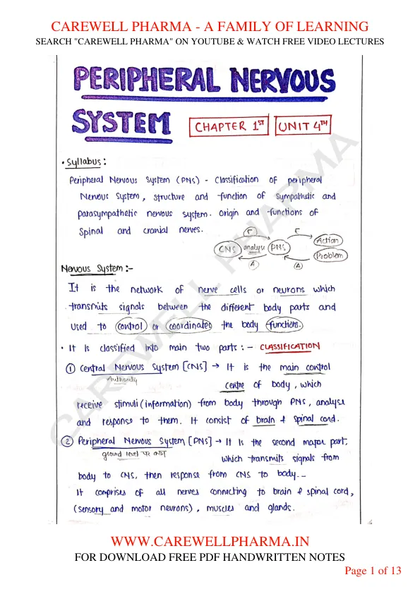
Syllabus : Peripheral Nervous System (PNS) Classification of peripheral Nervous system, structure and function of sympathetic and Parasympathetic nervous system. Origin and functions of Spinal and cranial nerves.

Nervous System : It is the network of nerve cells or neurons which transmits signals between the different body parts and Used to control or coordinates the body functions.
- CLASSIFICATION: It is classified into main two parts: -
Central Nervous system [CNS] : It is the main control Centre of body, which receive stimuli (information) from body through PNS, analyse and response to them. It consist of brain & spinal cord.
Peripheral Nervous System [PNS] : It is the second major part, which transmits signals from body to CNS, then response from CNS to body. It comprises of all nerves connecting to brain & spinal cord, (sensory and motor neurons), muscles and glands.

Somatic Nervous System :
It is a voluntary responses of our body, which we can control itself. It contain both sensory and motor neuron, in which stimuli receive through sensory neurons and response through motor neuron. eg movement of limbs etc..
Autonomic Nervous System :
It is an involuntary responses of our body, which we can not control itself.
- eg heartbeat, digestion etc...
- It also contain both sensory and motor neurons (muscles, glands).
Sensory Neurons - It carry information from the neurons of body to the CNS of body.
Motor Neurons - They conduct nerve impulse from CNS to the smooth & cardiac muscles and glands.
- It is further divided into two types:-
1. Sympathetic Nervous System [SNS] :
These are those system which activated during abnormal condition and helps body to maintain homeostasis and survive in these situations.
- It control physical or emotional stress due to work of exercise, pain and unfavourable living condition etc..
- This system mainly work on 3F (fear, fight and flight).

- The sympathetic nervous system arise from the cell bodies from the Thoracic (T1) to the Lumbar (L3) region of the spinal cord.
- This system is also known as thoracolumbar outflow.
- The primary neurotransmitter of SNS are dopamine, epinephrine and norepinephrine.
Functions
It performs various functions such as:
- release of sweat from sweat gland.
- contraction of arrector pili muscles.
- dilates pupils of iris.
- dilation of blood vessels of skeletal muscles.
- vasoconstriction of blood vessels of skin/mucous membranes.
- decreases the digestion rate.
- increases the heart rate & contractility, and raises BP.
2. Parasympathetic Nervous System [PSNS] :
These are those system which helps body to get back into normal condition after any abnormal situation.
- This system is mainly work for Rest and Digest.
STRUCTURE :
- the fibres of PSNS arises from the cell bodies of the CNS.
- The preganglionic fibres originated from the nuclei of the third, seventh, ninth and tenth cranial nerves (III, VII, IX, X) and the fourth sacral segments (S2 to S4) of the spinal cord.
- This system is also known as croniosacral outflow.
- The primary neurotransmitters of PSNS are acetylcholine (Ach) and peptides (such as cholecystokinin).

Functions
- It constricts pupils
- It facilitates flow of tears
- It facilitates flow of secretion of saliva.
- decreases rate of heart beat (BP $\downarrow$)
- Normalise/increases digestion rate.
- stimulates erection.
Differences between SNS and PSNS
| Sympathetic Nervous system | Parasympathetic Nervous System |
|---|---|
| Involved in the fear, flight or fight responses. or activates in abnormal condition. | Involved in maintaining homeostasis and permits the rest and digest response. |
| prepare the body for any potential danger. | to bring the body to a state of calm. |
| originated from all thoracic and upper 3 lumber | originated from cranial 3,7,9,10 and sacral 2,3,4 segments. |
| increases heartbeat, muscle tense up | reduces heartbeat, muscle relaxes. |
| pupil dilates | pupil contracts. |
| Saliva secretion is inhibited | saliva secretion increases |
| digestion rate decreases | digestion rate increases |
| releases epinephrine (adrenaline) and norepinephrine (nor-adrenaline) | releases acetylcholine |
- These both system work together to maintain homeostasis or equilibrium of our body.
Cranial Nerves
These are those nerves which originated from the brain and brainstem and extend throughout the body on both sides.
- Cranial nerves carry information from the brain to the other parts of the body, mainly to the head and neck.
- These nerves are present in paired.
- They are mainly responsible for facilitating smell, vision, hearing and movement of muscles.
- Twelve pairs (12) of cranial nerves are present in humans.

I - Olfactory
II - Optic
III - Oculomotor
IV - Trochlear (pathetic)
V - Trigeminal / Trifacial (Trigeminus)
VI - Abducens
VII - Facial (portio dura)
VIII - Auditory (portio mollis) / Vestibulocochlear
IX - Glossopharyngeal
X - Vagus (pneumogastric)
XI - Accessory / Cranial accessory XII - Hypoglossal
SPECIAL SENSES
CHAPTER 2
UNIT 4
- These are those sense organs which receive stimuli and convert them into nerve impulses.
- These impulses carry afferent or sensory nerve fibres which send information to the brain/CNS.
- These organs helps our body to perform various Important functions.
Examples
- Eye - for vision/sight
- Ear - for hearing
- Tongue - to sense the taste
- Nose - to sense the smell.
- Skin - to sense touch, pressure, temperature. (already done)
HUMAN EYE
- It is the highly specialised sense organ present in human body and responsible for the vision/sight.
- Eyes are almost spherical in shape having a diameter of about 2.5 cm. (25mm)
- They are situated in the orbital cavity (bony socket) in which bony wall protects eye from any type of injury.
- Both the eyes are coordinated in such a manner that they function as pair.

- It is made up with a various types of muscles and tissue.
It's structure can be classified into two parts:-
- External structure
- Sclera
- Cornea
- Iris
- Pupil
- Ciliary body
- Lens
- Internal structure
- Aqueous humour
- Retina
- Optic nerve
- Vitreous humour
- Choroid
ACCESORY STRUCTURE OF EYE
These are those structure which provide protection and support to the eye.
Eye brows: These are thick hair present over the eyes, which prevents the eye from sweat, dust or any other foreign particles from getting into the eyes.
Eye lids: They are the upper and lower folds of skin which protects the eye and provide closing and opening of eye.
At edge of eyelids, small hairs are present known as eyelashes.
Lacrimal gland: Those glands which involved in the production and secretion of tears.
Tears have antibacterial and lubricating properties, which clean the eyes & protected.
STRUCTURE OF EYEBALL
Sclera: It is a white visible portion of eye. It is made up of dense connective tissue and protects the inner part.
Cornea: It is transparent, anterior/front part of our eye, which covers the pupil and iris. the main function of cornea is to focus the light rays on retina.
Conjuctiva: It lines the sclera and inner part of eyelids. It keeps our eyes moist and clear and provide lubrication.
Iris: It is the pigmented, coloured portion of the eye, visible externally.
- The main function of the iris is to control the diameter/size of the pupil, according to light source.
- In low light size of pupil increased to get more light.
Pupil: It is the small aperture / hole located in the centre of the iris. It allow lights to enter and focus on the retina.
Lens: It is a transparent, biconvex lens of eye. It is present behind the pupil.
- It is attached to the cilliary body by ligaments.
- The lens along with Cornea refracts light/image so that it focuses on the retina.
Ciliary body: It is anterior part of choroid, which convert ciliary muscles and controls the shape of lens.
- Also, it helps in the production of aqueous humor.
Choroids: It is located between the sclera and retina and contain blood supply.
Retina: It is the innermost layer of eye. It is light sensitive and act as a film of a camera.
- Three layer of neural cells are present in them.
- Ganglion, bipolar and photoreceptor cells (rods for dark vision and cones cells for day vision).
- The main function of retina is to receive light through lens and formed image on retina, then convert this into electric nerve impulse for visual perception by the brain.
Optic nerve: It is located at the posterior portion of the eyes.
- It carry all the nerve impulse from the retina to the human brain for image perception.
Aqueous humour: It is a watery fluids present between the cornea and the lens. It nourishes the eye and keeps it inflated. (फूला)
Vitreous humour: It is a transparent, jelly-like substance present between the lens and the retina.
- It contains water (99%), collagen, proteins etc.
- The main function is to protects the eye and maintain its shape.
PHYSIOLOGY OF VISION
Light rays from an object enters into eye through cornea. $\downarrow$ Now, light passes through aq. humour and enter into pupil ( size of pupil adjusted by iris) $\downarrow$ Now, light enter into lens and lens focus the light rays on to the retina $\downarrow$ then, retina convert into nerve impulse, image (on the retina) $\downarrow$ which send to the brain through optic nerves. $\downarrow$ finally, seen by brain as Visual image

DISORDERS
Sty: In this, the margin of the eyelid get swelled up having a sensation of pain.
Conjunctivitis: In this, conjunctiva gets inflammed. It may occurs due to bacterial/viral infection or allergy. Madras/pink eye.
Color blindness: not clear vision, due to heredity, optic nerve or retinal disease.
Night blindness: difficulty in vision in night (low light). It may be due to deficiency of retinol (vitamin A).
Myopia: difficulty in seeing the distant objects.
Hypermetropia: difficulty in seeing the near objects.