Human Anatomy And Physiology 1 - Unit 3
Syllabus
Body fluids and blood
Body fluids, composition and functions of blood, hemopoeisis, formation of hemoglobin, anemia, mechanisms of coagulation, blood grouping, Rh factors, transfusion, its significance and disorders of blood, Reticulo endothelial system.
Lymphatic system
Lymphatic organs and tissues, lymphatic vessels, lymph circulation and functions of lymphatic system.
Scroll to Download
BODY FLUIDS & BLOOD
UNIT 3
CHAPTER 1
SYLLABUS : Body fluids, composition and functions of Blood, Hemopoeisis, formation of Hemoglobin, Anemia mechanism of coagulation, Blood grouping, Rh factors, Transfusion, its significance, Disorder of Blood, Reticulo Endothelial system.
Body fluids : These are those fluids which are present inside human body. About 50-60% of human body is fluids approx. (vary from person to person).
Based on distribution, fluids are distributed into two types:-
Intracellular fluids (ICF) The fluids which are present inside the cells, it is about 40-45% of body weight.
Extracellular fluids (ECF) The fluids which are present outside the cells, It is about 15-20% of body weight.
- Interstitial fluids - which is present between the cells (12-15%).
- Plasma - which is present inside vascular system (blood) - (4-5%).
- Others - Lymph, cerebrospinal fluids, synovial fluids, eye fluids etc.

BLOOD
A Blood is a liquid connective tissue in the body that flow through the vessels to act as a medium for the transport of oxygen, nutrient and hormones.
- The main role of blood is to transport the oxygen from lungs to all over the body and to protects the body against disease.
- The branch of medical science that deals with the study of blood is known as Haematology.
Properties of Blood:
- It is red color, slightly viscous fluid.
- It's pH is slightly alkaline i.e. 7.35 - 7.45 (pH range).
- In males, the volume is about approx. 5-6 litre.
- In females, the volume is about approx. 4-5 litre.
- Around 8% of total body.
FUNCTIONS OF BLOOD
Blood has mainly three functions
- Transport
- Regulation
- Protection
- Transport -
- It transport to the cells from the lungs and from cells to the lungs.
- It transport nutrients from the digestive organs to the cells.
- It transport hormones and enzymes to the target tissue and glands.
- It transport waste products from body cells to kidney, lungs and sweat glands.
- Regulation -
- It regulates pH of our body through buffers.
- It regulates body temp through the heat absorbing and coolant properties of its water constant.
- It regulates water balance.
- Protection -
- protects body against blood loss through its blood clotting mechanism.
- It protects body against disease/pathogens through phagocytes, WBC (white blood cells) and antibodies.
COMPOSITION OF BLOOD:
It consist of blood plasma and formed element.

Blood Plasma : It is liquid part of blood, which act as a solvent for blood. It is pale yellow colored and consist 55% of total blood.
It contain several components -
- Water : It is the major part of blood (about 90% of plasma).
- It maintain blood pressure and takes part in body fluid level and electrolytes.
- Proteins: maintain osmotic pressure
- Albumin, takes part in immunity and inflammation
- Globulin, takes part in defence and in blood coagulation
- Fibrinogen, contain various blood clotting factors.
- Others: contains nutrients (glucose, lipids, vitamins, electrolytes, amino acids) and solutes like , , , etc.
FORMED ELEMENTS : Also known as Blood cells. It is about 45% of total Blood. It is composed of RBCs, WBC, and Platelets.
- Red Blood Cells (RBC) : Also known as Erythrocytes.
- These are disc-shaped cell and visible under microscope.
- Functions: It contain a pigment called Hemoglobin ($Hb$) which transport all over the body.
- The total life span of RBCs is about 120 days.
- RBCs are produced in spleen and bone marrow.
- The total counts of RBCs is about 5.4 million per in males and 4.8 million/$\mu l$ in females.
- White Blood Cells (WBCs) : Also known as Leukocytes.
- They are round or irregular, semi-transported cells, containing a nucleus and visible under microscope.
- Functions: They fight against infections/pathogens and protects us from disease. (soldiers of Body defence system).
- WBCs act as a Body's defence system, it make some chemical entity called antibodies to fight against infections.
- They have larger size than RBCs but low in quantity, the total count of WBCs are 4500 - 11000 per (microlitres).
- It contains:
- Granulocytes
- Agranulocytes
Granulocytes: they contains granules.
- Neutrophils - It is about 60-70% of total WBCs. It attack the invading bacteria and engulf them.
- Basophils - It is about 0.5-1% of total WBCs. they release heparin which prevent blood clotting and histamine which promotes inflammation.
- Eosinophils - It is about 2-4% of total WBCs. they play a major role in immune system & reduce inflammation.
Agranulocytes: not contain granules.
- Lymphocytes - It is about 20-25% of total WBCs. they produces antibodies which protects body and also provide immunity.
- Monocytes - It is about 3-5% of total WBCs. they work as defence mechanism of body.
- Blood Platelets : Also known as Thrombocytes.
- These are tiny granular cells, which formed in bone marrow, and they are surrounded by plasma membrane.
- A normal platelet count ranges from 150,000 - 450,000 Platelets per microliter ($\mu l$) of blood.
- The main role of platelet is to releases blood clotting factors and helps in blood coagulation (haemostasis). Also helps in defence system.
HAEMOPOIESIS
It is the process of formation of Blood cells (RBCs, WBCs, platelets).
- Haemopoiesis = Haemo/Hemo (Blood cells) + Poiesis (formation/development)
- The sites where this process occurs are hemopoietic tissues or organs (bone marrow, liver and spleen).
- In a person, approximately 200 billion new RBCs, 20 billion new WBCs and 25 billion platelets are formed each day.
Formation of Blood Cells
- Formation of blood cells occurs outside the bloodstream, and the newly formed cells into the circulation via diapedesis.

- Diapedesis - transport of blood cells into blood circulation.
- Blood formation occurs at different sites in different stages of life.
- In the earlier embryonic stage, blood cells develop from the mesoderm of the yolk sac (during pregnancy) - till approx 4 weeks.
- After 1-2 month, foetus gets mature and haemopoiesis starts in liver and spleen.
- After 2-3 month of pregnancy, haemopoiesis starts in bone marrow and continues even after birth.
- Infants and childrens have only red bone marrow, which further converts into yellow bone marrow as child grow.
- Thus in adults, the main site for hemopoiesis are red bone marrow, presents at the ends of long bones and flat bones (pelvis, ribs, sternum, scapulae & skull etc).
So, All the blood cells develop in the bone marrow from haemopoietic stem cells.
It is mainly of three types:-
- Erythropoiesis formation of Erythrocytes (RBCs)
- Leukopoiesis formation of Leukocytes (WBCs)
- Thrombopoiesis formation of platelets (thrombocytes).


- Erythropoiesis: It is the process of formation of Red blood cells (RBCs) or erythrocytes.
- It starts when a Haemopoietic stem cells generate myeloid progenitor cells.
- Then, with the help of erythropoietin (EPO) and other growth factor, it produced proerythroblasts (pronormoblasts).
- Then this gives small daughter cells i.e. erythroblasts or normoblasts which forms reticulocytes by loosing nuclei.
- At lasts, then this reticulocytes transport into blood circulation and mature into one to two days, into erythrocytes (RBCs).
- Thrombopoiesis: It is the process of formation of platelets.
- Megakaryoblasts are the precursor cells of platelets, which form megakaryocytes.
- Now, maturation of megakaryocytes are take place in bone marrow with the help of thrombopoietin (TPO) and forms platelets.
- Leukopoiesis: It is the process of formation of white blood cells or leukocytes.
FORMATION OF HEMOGLOBIN
- Hemoglobin (Hb) is a respiratory pigment present in Red blood cells (RBCs) and giving them red color.
- The main role of Hemoglobin is to carry Oxygen ($O_2$).
- It is synthesized during erythropoiesis (during formation of Red blood cells) within immature erythrocytes in red bone marrow.
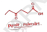
Structure : It consist of 4% heme ($Fe^{2+}$ and porphyrin) and 96% globin protein.
- Heme: It is a cyclic tetrapyrrole with four pyrrole molecules and responsible for red color of RBCs. It also contain iron ($Fe^{2+}$) atom.
- Globin: these are protein chain, which cover iron atom.
- Each molecules of Hemoglobin (Hb) consist of four globin chains i.e. two ($\alpha_1 + \alpha_2$) and two ($\beta_1 + \beta_2$) - these four chains form two dimers, i.e. and .

Properties: Normal hemoglobin level in
- males is 14-17 gm/100ml
- females is 12-15 gm/100ml
- and In newly born babies is 14.5 - 18.5 gm/100ml.
Synthesis of Hemoglobin:
- The synthesis of haemoglobin is initiated during erythropoiesis in mitochondrion.
- It involve several steps:-
The succinyl-CoA and glycine condense to form 5-aminolevulinic acid (pyrrole molecules). $$2 \text{ succinyl-CoA} + 2 \text{ glycine} \rightarrow
Four pyrrole molecules form protoporphyrin IX by combining together.
Now, protoporphyrin IX forms the heme molecules by combining with iron. $$Protoporphyrin \ IX + Fe^{2+} \longrightarrow Heme$$
Now, each heme molecules combined with globin (a long polypeptide chain synthesized by ribosomes). $$Heme + polypeptide \ globin \rightarrow Hemoglobin \ chain$$
Four of these chains loosely combine with each other, forming a hemoglobin molecules. $$2\alpha \ chains + 2\beta \ chains \rightarrow Hemoglobin$$

Functions/Importance : The main role of Hemoglobin (Hb) is to carry (Oxygen) from lungs to all over the body and transport back into lungs.
ANAEMIA
- It is defined as it is the reduction of Hemoglobin or Red Blood Cells (RBCs) concentration of blood below normal range.
- Due to deficiency of blood (RBCs), there will be decreasement in the amount of Oxygen ($O_2$) in body, which can cause serious damage to our body.
Causes/Reason: It occurs due to:
- excessive blood loss
- increased breakdown of RBCs
- defected RBCs production
- Nutritional defects
Symptoms: Pale skin, Weakness, Tiredness, Shortness of breath, fast heartbeat, Chest pain.
Types of Anemia:
- Iron deficiency Anaemia - It is the most common type of anemia in the world. It is occurs due to deficiency of Iron ($Fe^{2+}$) atom in blood. due to excessive loss of blood or due to low consumption of iron rich foods.
- Megaloblastic Anaemia - It is occurs due to deficiency of folic acid (folate) which are used in the production of RBCs. Due to this, it forms immature RBCs (megaloblasts) large erythrocytes. It occurs due to Insufficient diet or disease.
- Pernicious anaemia - It is occurs due to deficiency of Vit . It is an autoimmune disease, i.e. auto-antibodies damage the Intrinsic factor (IF).
- Hypoplastic and Aplastic anaemia - these anaemia occurs due to bone-marrow disease.
- Hypoplastic anaemia occurs due to decreased marrow function.
- Aplastic anaemia occurs due to destruction of bone marrow.
- Haemolytic anaemia - Abnormal condition, in which Red blood cells (RBCs) are damaged or removed from the blood circulation.
- Sickle-cell anaemia - It is comes under haemolytic anaemia, in which abnormal shaped red blood cells which may cause blockage of blood flow.
HAEMOSTASIS
- It is also known as Blood Coagulation or clotting.
- It is a process (sequence of responses) that stop Bleeding and reduces blood loss when blood vessels are damaged or ruptured.
- It is important to stop bleeding because excessive blood loss can develop deficiency of Oxygen ($O_2$) in body.
- This process involves various clotting factors and at last generate fibrin which forms clots (network of fibrins).
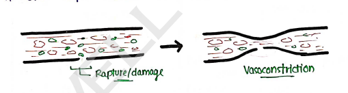
Mechanism of Blood coagulation/clotting : Hemostasis includes three steps that occurs in blood coagulation:
- Vascular Spasm
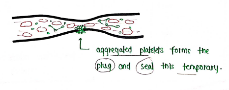
- Formation of platelet plug
- Blood coagulation / clotting
Vascular Spasm : It involves vasoconstriction in which the smooth muscles of blood vessels wall contracts which slow blood loss (narrow vessels pass less blood as compared to wide blood vessels). It also helps in the activation of platelets.

Formation of platelet plug In this process platelets aggregates and formed plug which sticks to the damage blood vessels and prevents temporary blood loss. aggregated platelets forms the plug and seal this temporary.
Blood coagulation / clotting In this process, fibrins is formed which are insoluble protein which further forms clots (network of fibrins). Now, these clots trapped RBCs and seal the damaged blood vessels. This process involves thirteen factors and three pathways.

Blood clotting factors The following table:
| No. | Name | Pathways | Sources |
|---|---|---|---|
| I | Fibrinogen | Common | Liver |
| II | Prothrombin | Common | Liver |
| III | Tissue factor (TF) or Thromboplastin | Extrinsic | Damaged tissue and platelets |
| IV | Calcium ion ($Ca^{2+}$) | All | Bones and Diet |
| V | Proaccelerin | Extrinsic & Intrinsic | Liver & Platelets |
| VII | Proconvertin | Extrinsic | Liver |
| VIII | Antihemophilic factor | Intrinsic | Liver |
| IX | Christmas factor (PTC) | Intrinsic | Liver |
| X | Stuart-Power factor, Thrombokinase | Extrinsic and Intrinsic | Liver |
| XI | Plasma thromboplastin antecedent (PTA) | Intrinsic | Liver |
| XII | Hageman factor | Intrinsic | Liver |
| XIII | Fibrin stabilizing factors |
It involve three stages
- Formation of prothrombinase.
- Conversion of prothrombin into thrombin.
- Conversion of fibrinogen into fibrin.
Mechanism of Blood Coagulation

Formation of Prothrombinase: It involves both extrinsic and intrinsic pathway in which both sense trauma and starts the process of coagulation. By the sequence of reaction or by activating many clotting factors, both pathway forms prothrombinase enzymes (shown in flowchart).
Conversion of prothrombin into thrombin: It involves common pathway in which prothrombin (factor II) convert into thrombin (IIa) with the help of enzyme prothrombinase and ions.
Conversion of fibrinogen into fibrins: It also involves common pathway in which fibrinogen (protein present in plasma) convert into fibrin threads, with the help of thrombin. Then these fibrin forms network of fibrins i.e. clots, then these clots trapped RBCs and seal the damaged blood vessels.
BLOOD GROUPING
- It is the classification of blood, on the basis of the presence of antigens on the surface of RBCs.
- It is important for blood transfusion i.e. transfer/replacement of blood cells. (essential to know blood group of both donor and receiver).
- It helps in diagnosis of some disease (hemolytic disease).
- There are two main blood group system, which are responsible for people's blood group.
- ABO Blood Group
- Rh system
1. ABO Blood Group System: It is most important system which is based on A and B antigens on the surface of RBCs, Plasma containing circulating antibodies (a & b) in plasma. ABO blood types were discovered by Karl Landsteiner.


Type A: It consists of protein A antigen which present on the surface of RBCs. It also contain antibodies b. Blood group A person can receive blood from Group A and O and can donate blood to Group A & AB.
Type B: It consists of Antigen-B present on the surface of RBCs and (anti-a) antibodies. Blood group B person can receive blood from Group B and group O, and can donate blood to group B & AB.
Blood group Type AB: It consists of Antigen A and Antigen B both but it contain no antibodies. It is Universal receptor which can receive blood from any blood group person. It can donate its blood to only blood group AB.
Blood group type O: It does not consist any Antigen but it contain both (anti-a & anti-b) antibodies that's why they have strong immunity. It is universal donor which can donate blood to anyone, but it can receive blood from only group O.
2. Rh Blood Group System: This system is mainly used for the identification of presence and absence of Rh antigen on the surface of RBC.

- Rh positive (+ve): If Rh antigen present on surface of RBCs then it is positive (+ve). (more than 90%).
- Rh negative (-ve): If Rh antigen is absent on the surface of RBC, then it is negative (-ve). It was first detected in Rhesus monkey, so it is called Rh factor. Incompatibility b/w (+)ve & (-)ve: Erythroblastosis foetalis which cause foetal death in the womb.
BLOOD TRANSFUSION
It is a life saving procedure, which is used for replacement of blood cells, in deficiency or loss of large amount of blood through bleeding.
- It becomes essential in condition like anaemia, haemorrhage, trauma, burns and surgery.
- Donor: A person who donates the blood.
- Recipient: A person who receives the blood.
- It is performed only in case of emergency/critical condition. Cross-matching should always be performed.
Significance of Blood Transfusion It is used in condition of -
- Blood loss
- Blood disease
- Acute poisoning
- Hemolytic disease.
DISORDERS OF BLOOD
There are many disorder and blood related disease.
- Anemia deficiency of RBCs or hemoglobin in blood.
- Leukaemia (Blood cancer) It is a condition of cancer which is characterised by multiplication of abnormal WBCs in uncontrolled manner.
- Thrombocytopenia It is the low level of blood platelets.
- Polycythaemia It is a condition having a high concentration of red blood cells in blood.
- Coagulation disorder uncontrolled bleeding, even in a minor trauma.
RETICULOENDOTHELIAL SYSTEM
It is also known as RES, Mononuclear-Phagocytic System (MPS) and Tissue-macrophage system.
- It is one of the defensive system of our body in which phagocytic cells prevents from foreign substances or invading bacterias by engulfing them.
- From the bone marrow the monocytes enter to blood circulation and remain for three days and then migrate into the tissues.
- In tissues, these cells attain maturity, develop phagocytic property and finally converts into macrophages.
- These tissue macrophages are spread to different parts of body and present at the sites of:- Bone marrow, Lymph nodes, Lungs, liver, spleen, bones etc.
Functions:
- Protect against bacteria invading the body tissue.
- Play a role in inflammation and healing.
- Boost up the immunity/immune responses.
- Remove old RBCs, WBCs and platelets.
LYMPHATIC SYSTEM
UNIT 3
CHAPTER 2
- Lymphatic system is a closed network of lymphatic vessels through which (lymph) circulates throughout the body and protects body against harmful agents and maintain bodys fluid balance.
- The flow of lymphatic fluid is undirectional i.e. lymph flow from tissue spaces to the blood.
LYMPH
- It is a clear watery fluids that flow through vessels.
- The major functions of lymph is to transportation of plasma proteins, back to the bloodstream and Nutrition & oxygen is supplied to low blood areas.
Functions
- Drainage - maintain constant volume and composition of tissue fluids by removing excess fluids and metabolites.
- It also absorb small intestinal fats through lymphatic capillaries i.e. lacteals (it look like milky).

FORMATION OF LYMPH & CIRCULATION OF LYMPH
As we know, blood flow throughout the body through blood vessels. These blood vessels divides into small-small blood capillaries, through which blood transport oxygen & other nutrients to all over the cells.

PARTS OF LYMPHATIC SYSTEM It consists of :-
- Lymphatic vessels / capillaries which contain lymph and flow inside it.
- Lymph
- Lymph Organs
- Bone marrow: produce mature B-Lymphocytes (production of blood).
- Thymus: mature T-lymphocytes.
- Secondary: Lymph nodes which filter lymph.
- Spleen: used for destruction of older RBC's.
LYMPH NODES :
Also known as Lymph glands or lymphatic nodes.
- These are small and glandular organs of lymphatic system which presents along the path of lymph vessels.
- The main function of lymph nodes is to filter or trap foreign particles, and trap cell debris, bacteria etc.
- They also multiplying lymphocytes by pre-existing lymphocytes, then pass into blood stream. Also produce $\gamma$-globulin.
- The lymphatic vessels consists of nearly 600 bean-shaped lymph nodes, usually distributed all over the body, and in large amounts under the shoulder joint (axilla), elbow, knee and groin (b/w upper thighs & lower abdomen).
STRUCTURE :
It consist of three parts:-
- Cortex It is the outer zone of densely packed lymphocytes, from which lymph enters.
- Paracortex It is present between the cortex and medulla and comprises of T lymphocytes.
- Medulla It is densely occupied by macrophages, the B cells and the antibody producing plasma cells, migrated from cortex into medulla.

FUNCTIONS OF LYMPHATIC SYSTEM
- Tissue Drainage: Interstitial fluids/lymphatic system return blood back to circulation, other lymph go back to veins. (tissue to blood).
- Immunity: lymphatic system contains mainly lymphocytes (B & T) which provides immunity. Lymphocytes & monocytes are body's defence cells that remove bacteria or foreign agents.
- Absorption: Lymphatic vessels lacteals, that absorb fats from small intestine and also fat soluble vitamines (Vit. A, D, E & K).
